Humanized Strategy
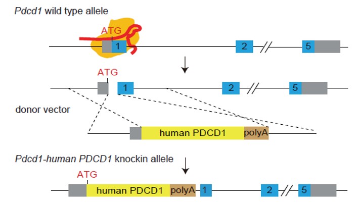
Strategy for Generating Humanized PD-1 (ASHU-00015) Mouse Model
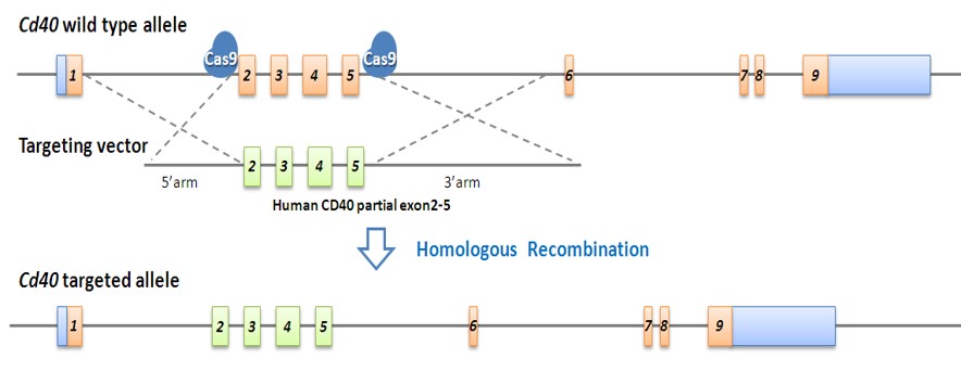
Strategy for Generating Humanized CD40 (ASHU-00076) Mouse Model
Validation Data: PD-1
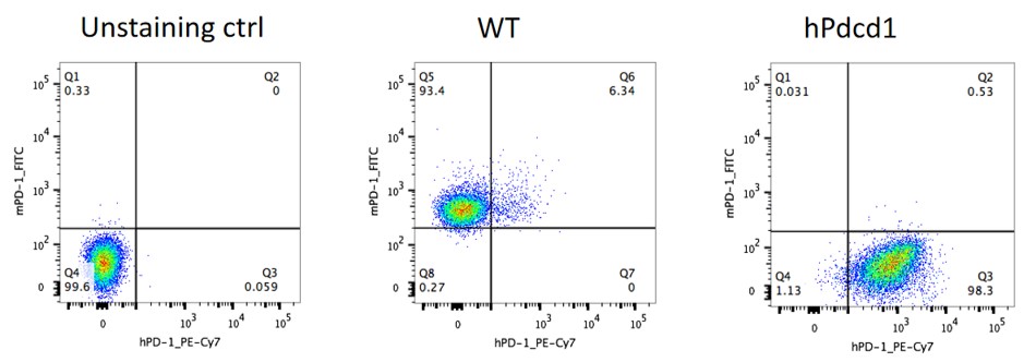
Figure 1. Expression of PD-1 in the activated spleen lymphocytes of humanized PD-1 Homozygous mice is detected by FACS.
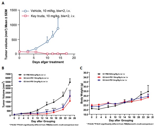
Figure 2. In vivo anti-tumor effect of an anti-human PD-1 antibody in a humanized mouse model of PD-1. Anti-human-PD-1 drugs significantly inhibited the growth of MC38 tumors in PD-1 mice, demonstrating that the humanized PD-1 mice can be used to assess the anti-human PD-1 antibody.
A. Mean volume ± SEM of tumor tissues (Data in partnership with collaborators) In vivo validation of anti-tumor efficacy in a MC38 tumor-bearing model of humanized PD-1 mice. Homozygous humanized PD-1 mice were inoculated with MC38 colon cancer cells. After the tumors grew to 100 mm3, the animals were randomly assigned into a control group and a treatment group (n=8). The drug was given twice a week for a total of 4 administrations. The results showed that Keytruda, a drug targeting human PD-1, exerted a very significant anti-tumor effect (p<0.001), demonstrating that the humanized PD-1 mice are a good in vivo model for validating the efficacy of antibodies targeting human PD-1.
B. Mean volume ± SEM of tumor tissues. C. Mean body weight ± SEM of mice (Data in partnership with collaborators) In vivo dose validation of anti-tumor efficacy in a MC38 tumor-bearing model of humanized PD-1 mice. Homozygous humanized PD-1 mice were inoculated with MC38 colon cancer cells. After the tumors grew to about 90 mm3, the animals were randomly assigned into a control group and a treatment group (n=9). The results showed that the antibodies targeting human PD-1 showed a very significant antitumor effect (p<0.001), and such antitumor effect is dose-dependent.
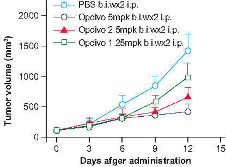
Figure 3. In vivo dose validation in a MC38 tumor-bearing model of humanized PD-1 mice. Homozygous humanized PD-1 mice were inoculated with MC38 colon cancer cells. After the tumors grew to 100 mm3, the animals were randomly assigned into a control group and a treatment group (n=8). The drug was given twice a week for a total of 4 administrations.
Validation Data: CD40
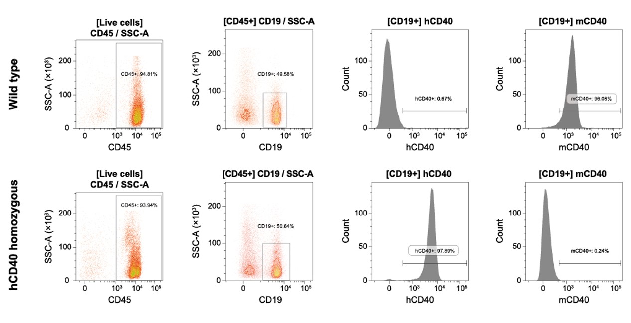
Figure 4. Detection of CD40 expression in peripheral blood cells of humanized CD40 mice. The FACS results of peripheral blood cells collected from homozygous humanized CD40 mice and wild-type mice showed that the active expression of humanized CD40 was detected in CD19 positive cells collected from homozygous humanized CD40 mice, and its expression level was similar to that of murine Cd40 expression in wild-type mice. (Data in partnership with collaborators).
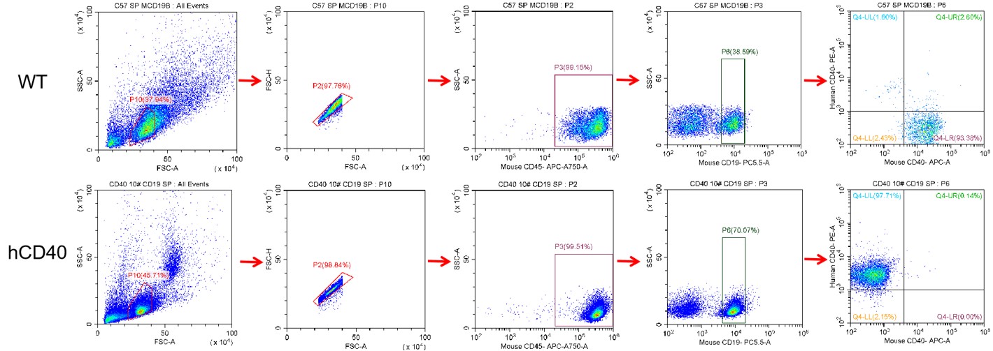
Figure 5. Analysis of human CD40 expression in the spleen by FACS. The homozygous KI animal expresses human CD40 on the CD19+ B cells.
Reviews
There are no reviews yet.