
The endogenous mouse Cd3e/Cd3d/Cd3g genes were replaced by human CD3E/CD3D/CD3G gene.
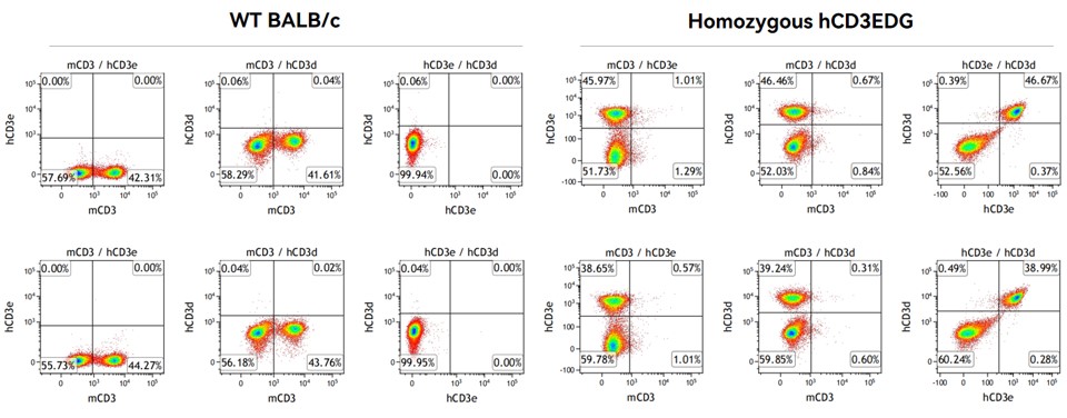
Figure 1. Detection of surface expression of human CD3E/CD3D on T cells in spleen in homozygous hCD3EDG (BALB/c) mice. (Data in partnership with collaborators)
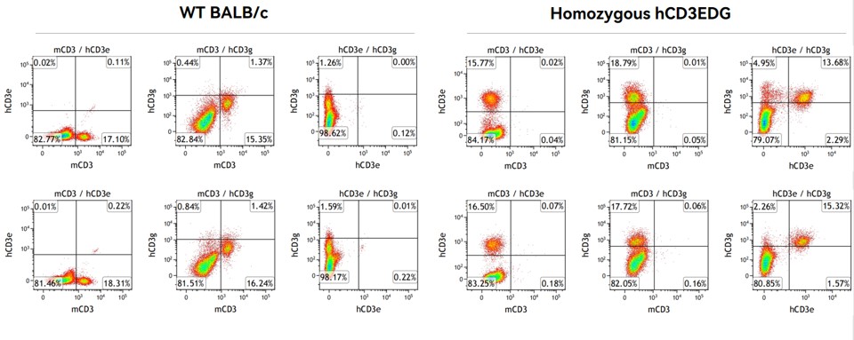
Figure 2. Detection of surface expression of human CD3E/CD3G on T cells in blood in homozygous hCD3EDG (BALB/c) mice. (Data in partnership with collaborators)
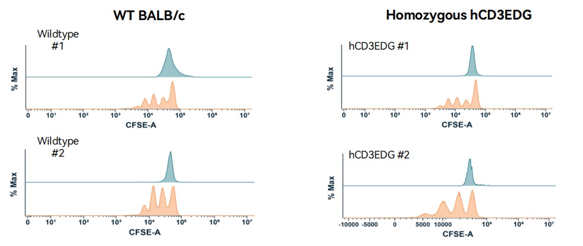
Figure 3. Analysis of TCR signalling in hCD3EDG (BALB/c) mice upon CD3/CD28 activation in vitro. (Data in partnership with collaborators)
Note: CFSE-dilution as a measure of mouse T cell proliferation.
CD3+ T cells were isolated in the splenocytes from WT BALB/c and hCD3EDG mice. Isolated T cells were labeled with µCFSE dye and stimulated with anti-mCD3 (5µg/mL) or anti-hCD3 (5µg/mL) plus soluble anti-mCD28 (50µg/mL) for in vitro culture for 72 hr. T cell proliferation was analyzed by flow cytometry for CFSE dilution.