G-banding is a cytogenetics technique that helps determine the number and appearance of chromosomes by staining condensed metaphase chromosomes to produce a visible karyotype. When stained with Giemsa stain, the heterochromatic regions will present darker while the euchromatin sections will present light bands in G-banding. The chromosomes are stained in a distinctive pattern that allows the pairing of homologous chromosomes and is useful for verifying the genetic integrity of cells (primary cell and iPSCs) and for identifying structural abnormalities, through the photographic representation of the entire chromosome within the cell. It is well-known that long-term in vitro culture of cells and passaging introduces karyotypic/ chromosomal abnormalities in 20-50% of cells (e.g., insertions, deletions, duplications, translocations), and may lead to invasive cell populations that could affect cellular phenotype and integrity of results. This is especially critical for iPSCs where the karyotype of parental tissue needs to be maintained in an uncompromised manner as they are used as biorelevant models of the patient physiology/ health. Because of this, ASC recommends karyotyping iPSCs or ESCs at passages 10-15.
G-banding karyotyping is performed as routine quality control (QC) screening at various stages of iPSC or cell culture: before/ after cell derivation or reprogramming; before/ after CRISPR genome editing/ differentiation; for cell banking; or when cultures show unusual growth properties.
Applied StemCell offers high-resolution G-Banding and chromosome counting service with fast turnaround times for your cell line with a full report analyzed by an experienced cytogeneticist and with publication-quality images. We also offer other karyotyping and copy number variation (CNV) analysis services such as the array comparative genomic hybridization analysis (aCGH) or chromosomal microarray analysis (CMA) for the detection of small deletions and duplications, quantitative PCR (qPCR) for determining chromosomal integrity or abnormalities in your cell lines.

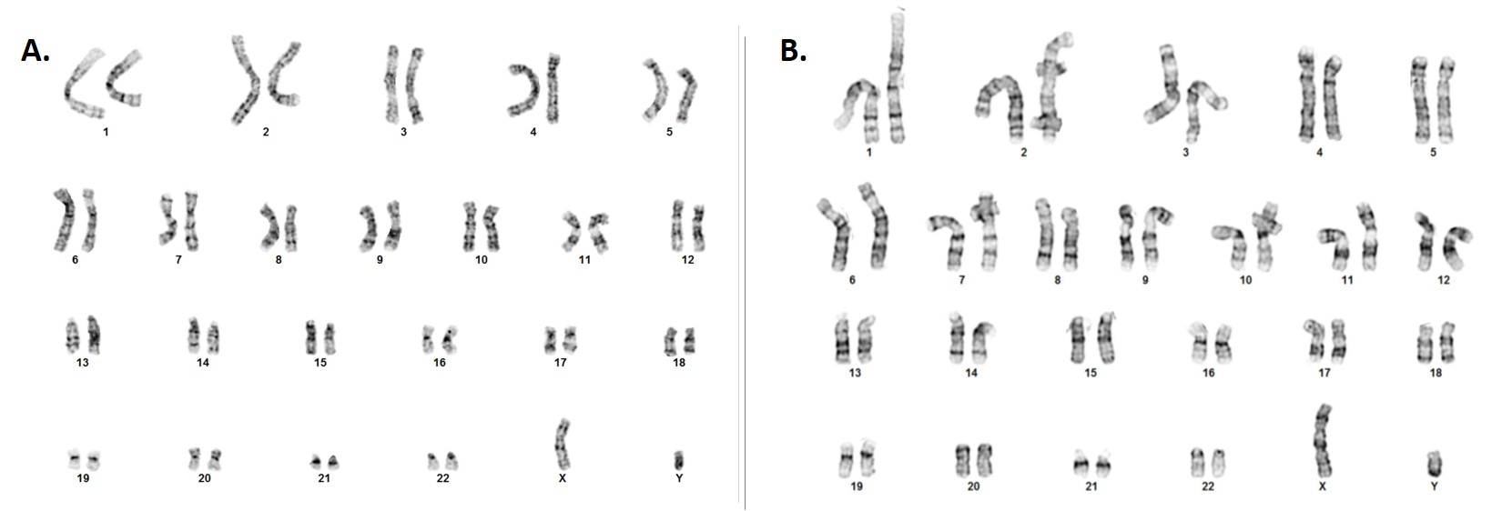
Figure. Representative G-banding karyotype images of control iPSC lines at passage 17 (p17) analyzed in two facilities: (A) a reputed, local institution that routinely offers cytogenetics services; (B) Applied StemCell’s (ASC) high resolution karyotyping service. Results from the local institution’s cytogenetics lab (image A) examined 20 metaphase cells and reported a normal male karyotype in all cells. Results from ASC’s cell line characterization of the same cell line at same passage (image B) but using a higher resolution technique reported that out of 20 metaphase cells examined, fifteen cells (15) reported an apparently normal karyotype, four (4) cells demonstrated what may be a small duplication of the long arm of chromosome 20 band q11.2 which includes the BCL2L1 gene and is associated with a moderate proliferative growth advantage in human pluripotent stem cells (PSCs) enabling a few cells with trisomy 20 to completely replace normal cells. One (1) cell demonstrated a non-clonal aberration.
Applied StemCell (ASC) offers several other karyotyping methods, including telomeric staining (T-banding), constitutive heterochromatin staining (C-banding), reverse Giemsa staining (R-banding), and quinacrine staining (Q-staining), so you can fully characterize your iPSCs to confidently move your research forward. If you do not see what you are looking for, contact us today to speak with one of our experts.